Secondary Glaucoma
'Secondary' means a form of Glaucoma the cause of which can be attributed to another underlying ophthalmic or medical condition that predisposes to the development of high eye pressure. This can be injury, disease or a side effect of medication.
Strictly speaking, the condition is called ‘secondary ocular hypertension’ if there is just raised intraocular pressure but with no detectable damage to the optic nerve. If, however, the raised pressure damages the optic nerve, which therefore can lead to visual loss, this would then be called secondary glaucoma.
The good news, in many cases, is that if the underlying cause is fixed, the eye returns to normal and further treatment is unnecessary. Unfortunately, damage to either the optic nerve or visual field cannot be reversed, but should stabilise once the pressure is normalised.
Causes of Secondary Glaucoma
1. Pigment dispersion syndrome and pseudoexfoliation
These are two of the commonest causes of secondary open angle glaucoma. Because they are treated in an identical fashion to primary open angle glaucoma, some doctors classify them as a version of the same condition.
In pigment dispersion syndrome, pigment granules are intermittently rubbed off the back of the iris as it moves and touches the lens and these granules become deposited in the trabecular meshwork. This causes a blockage to the drainage system which leads to high intraocular pressure. When it causes optic nerve damage it is termed pigmentary glaucoma. The condition is more common in young, myopic (short- sighted) men.
In pseudoexfoliation, the trabecular meshwork gets clogged up with white flakes. These flakes are deposits from the surface of the lens capsule, rather like dandruff, that get shed by the continuous movement of the iris (when the pupil gets larger or smaller). If pseudoexfoliation leads to damage to the optic nerve it is termed pseudo-exfoliative glaucoma. It tends to be more common with increasing age and is thought to be more prevalent in certain ethnic groups (such as those of Scandinavian and Southern Mediterranean descent), although can be seen in any ethnicity.
2. Iatrogenic
Iatrogenic literally means ‘caused by a doctor’. The main iatrogenic cause is following retinal surgery. This is because sometimes the retinal surgeon needs to put substances such as gas or silicone oil in the eye in order to flatten the retina following a retinal detachment or after other retinal surgical treatments. These substances invariably cause the pressure to rise and sometimes the rise in pressure can be very high. With gas, the effect is usually short-term and some of the gas may be removed early to reduce the pressure. Silicone oil may need to stay inside the eye for many months, years or indefinitely and a high proportion of these patients risk developing secondary glaucoma.
Another common cause for high pressure is the use of steroids – in the form of tablets, face creams, nasal sprays and, in some cases, extended use of steroid eye drops. Steroids are far from the only medication that can cause glaucoma and it is a complex area.
3. Inflammation in the eye (uveitic glaucoma)
Uveitis is inflammation of the layer of pigmented tissues inside the eye (uvea, made up of the iris, ciliary body, and choroid). There are a number of different ways in which uveitis can present – anterior uveitis or iritis will only affect the front of the eye; vitritis is inflammation in the vitreous; posterior uveitis is inflammation in the back of the eye.
Uveitis can cause leakage of protein and white blood cells into the aqueous, and these then deposit in the trabecular meshwork and block the drainage system. The trabecular meshwork itself can become inflamed ‘trabeculitis’, swelling to block the spaces in the drainage system through which aqueous normally leaves the eye. The current thinking amongst eye specialists is that this may increase pressure when the eye already has high pressure but only very minimal inflammation, such as the condition known as ‘Posner- Schlossman syndrome’ or when shingles affects the eye).
Uveitis may cause secondary angle closure. Inflammation in the front part of the eye can cause the iris to stick to other structures. Sometimes the iris around the pupil can stick to the lens (posterior synechiae); if the pupil gets completely stuck down for 360 degrees then there is no gap and aqueous cannot pass from the ciliary body to the anterior chamber. This means that aqueous builds up behind the iris, pushing it forwards and completely closing off drainage through the trabecular meshwork. This causes the pressure to go very high quickly and will require urgent intervention.
The pressure can be relieved by making a hole (iridectomy) in the iris to allow the aqueous to pass. This is preferably achieved using a surgical procedure, but laser can be used as a temporary measure in an emergency setting.
When uveitis occurs over a long period of time, the iris can stick to the trabecular meshwork (peripheral anterior synechiae) and if these adhesions are extensive there will be a significant reduction in aqueous outflow, leading to high pressure and glaucoma. Often the treatment for this will need to be surgical drainage.
When high pressure occurs during an acute episode of anterior uveitis, it is called ‘hypertensive uveitis’. A rare variant of this, when the pressure is very high but the inflammation is minimal, is called ‘Posner-Schlossman’ syndrome. In most cases, the episodes are short- lived and respond well to topical steroids to treat the inflammation and glaucoma medications (eye drops and occasionally Diamox tablets) to lower the eye pressure. The medications can usually be stopped once the episode has resolved. In some patients, the episodes can recur or eventually stop completely.
Fuch’s Heterochromic Cyclitis is a specific form of uveitis associated with loss of pigment from the iris of the eye, which becomes blue if previously brown. Symptoms are often absent in the early stages but raised pressure and cataract development may occur later and affect vision.
4. Lens-related problems
When a cataract is very advanced it can become swollen and block the flow of aqueous from the ciliary body through the pupil. This causes a build up of aqueous behind the iris, pushing the iris forward, and closing off drainage through the trabecular meshwork. This is termed ‘phacomorphic’ and is relieved by removing the cataract.
A similar ‘pupil block’ mechanism may occur if the lens within the eye is unstable due to weakness or loss of the ligaments that support the lens (known as zonules). This can happen following trauma and in patients with pseudoexfoliation (where the zonules are weaker than normal).
Very rarely, when a patient has advanced cataract, proteins from within the lens leak into the aqueous and causes high pressure in the same way as with uveitic glaucoma. This mechanism is termed ‘phacolytic’ and again will respond to removal of the cataract. Much more rarely, if there is a break in the capsule surrounding the lens (for example following an injury), leakage of lens material may cause a severe reaction in the eye causing a lot of inflammation and secondary raised pressure. This is termed ‘phacoanaphylaxis’ and requires removal of the cataract.
Cataract surgery itself can create increased intracocular pressure. Sometimes the artificial lens must be inserted in the anterior chamber, rather than the posterior chamber. These lenses may impede aqueous passage through the pupil causing a secondary angle closure. It is therefore essential that a peripheral iridectomy (a hole in the peripheral iris) is made when the lens is inserted. In these cases, the eye pressure can become very high if the peripheral iridectomy has not been made or closes off.

When the artificial lens is inserted behind the iris but in front of the capsule (in the ‘sulcus’), the lens can rub pigment off the back of the iris. These fragments block the trabecular meshwork, leading to high eye pressure.
Even patients who have cataract surgery where they do not have an artificial lens inserted are not immune. Not having a lens is called ‘aphakia’ and it can cause a secondary glaucoma, although the precise mechanism is unknown. This is the most common cause of secondary glaucoma in children.
5. Trauma
A blow to the eye or a penetrating injury is an event to take very seriously. Inflammation or bleeding (with blood cells clogging up the drainage system) or damage to the lens can quickly result in increased pressure in the eye.
A chemical injury (such as an alkali burn) can lead to severe inflammation of the cornea and uveitis, leading to secondary raised pressure
Hit hard enough and the drainage angle can be pushed backwards permanently (angle recession). This can lead to the development of high pressure a number of years after the injury, so it is vital that having suffered eye trauma you are carefully monitored for glaucoma for a long time.
6. Medication-related
The most common medicine-related cause of secondary raised intraocular pressure is called ‘steroid response’. Steroids are a very common form of medication widely used in everything from asthma to arthritis. They can be inhaled, taken orally, injected, used on the skin or taken locally in the eye.
Problems usually occur where the steroid is applied locally to the eye, either via injection or a steroid eye drop. This is usually when they are used to treat inflammation.
In people who already have narrow drainage angles, any eye drop which dilates the pupil may result in raised pressure by closing the angle further.
7. Neovascular (newly formed blood vessels)
In certain retinal conditions, such as proliferative diabetic retinopathy and central retinal vein occlusions, the poor blood supply within the eye may cause new blood vessels to grow onto the surface of the eye and into the drainage angle. If the new vessels become extensive and are present for a long time they can scar and completely close the drainage angle (synechial closure).
Often patients will already have poor vision in the affected eye due to the underlying retinal problem. However, if the pressure and the underlying retinal condition treated are not adequately treated quickly enough, the secondary glaucoma due to the new vessels (also called rubeotic glaucoma) can lead to severe optic nerve damage with a high risk of blindness.
8. Following corneal disease and corneal surgery
Secondary ocular hypertension can occur because of disorders of the cornea (the clear window at the front of the eye). Corneal infection, whether bacterial or viral, and corneal burns may cause inflammation in the eye (similar to uveitis) leading to raised intraocular pressure.
Patients who have undergone corneal grafts require topical eye drop steroids for many months to prevent the corneal graft from failing. And as mentioned above, steroid drops are a known cause of secondary glaucoma in some patients.
The more complicated the corneal problems, the more involved the surgery. There is always the risk of scarring adhesions (synechiae) between the iris and the trabecular meshwork (peripheral anterior synechiae); this also occurs in patients following multiple graft surgery. If the synechiae are extensive, the outflow of aqueous can be severely reduced, raising pressure. In many cases, glaucoma drainage surgery may be required.
Very rarely, patients with multiple failed corneal grafts may need to have an artificial keratoprosthesis (artificial corneas) inserted to allow them to see. Keratoprosthesis patients have a high risk of developing secondary glaucoma, with the additional problem that the pressure in eyes with artificial corneas cannot be measured.
9. Iridocornealendothelial (ICE) syndrome
Iridocornealendothelial (ICE) syndrome is a rare disorder that can cause glaucoma in adults aged 30-50 years, more commonly in women. It is unilateral (occurring in only one eye) and is related to an abnormality of one of the cell layers of the cornea. ICE syndrome is associated with swelling of the cornea and acquired defects of the iris.
High pressure is caused by the formation of adhesions of the iris to the trabecular meshwork (peripheral anterior synechiae) reducing the outflow of aqueous. It can be difficult to control the pressure in such eyes using drops, so treatment will often require the implantation of a glaucoma drainage device (tube surgery).
10. Bleeding inside the eye
As mentioned in the section on trauma, blood cells in the anterior chamber can clog up the drainage channels and lead to high pressure. Blood in the vitreous cavity (vitreous haemorrhage – often occurring in proliferative diabetic retinopathy) can cause raised pressure by a similar mechanism.
11. Raised Episceral Venous Pressure
This refers to conditions where there is raised pressure in the veins into which the aqueous drains. This can happen when there is an increase in pressure in the tissues of the eye socket (e.g. in the presence of an orbital tumour or thyroid eye disease), or abnormalities of the veins, or an abnormal link between the arteries and veins (a fistula). This high pressure restricts the outflow of aqueous and therefore causes high eye pressure.
12. Tumours
Occasionally tumours may develop in the eye. Tumours in the iris may involve the drainage angle and therefore reduce drainage outflow causing high eye pressure. A tumour in the ciliary body may push the peripheral iris forward, closing off the drainage angle. Large tumours in the back of the eye may push the lens forward, closing off aqueous passage through the pupil leading to a secondary pupillary block angle closure. Tumours may also cause new vessel formation.
Quality care in a relaxing surrounding
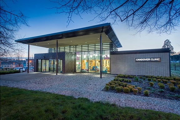
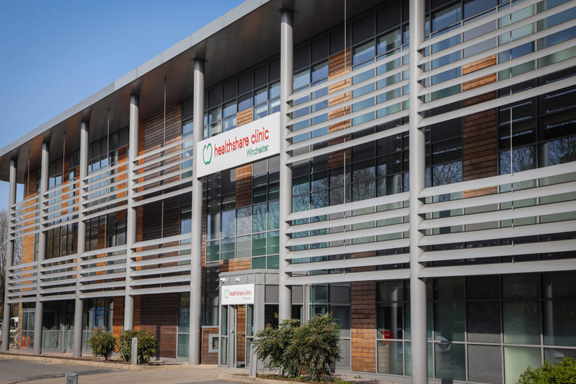
Book an appointment to see Mr Nish Srikantha at the Candover clinic in Basingstoke, or the Healthshare Clinic in Winchester.
Both locations are modern purpose built private hospitals designed entirely for patient well-being and comfort. The Candover Clinic is a stand-alone unit located on the Basingstoke and North Hampshire Hospital. The Healthshare Clinic on the outskirts of Winchester boasts a purpose built ophthalmic outpatients department and operating theatre offering the latest diagnostic and treatment technologies.